Dr Tricia Ruth Thomas, Obstetrics and Gynaecology (O&G) from Pantai Hospital Klang.
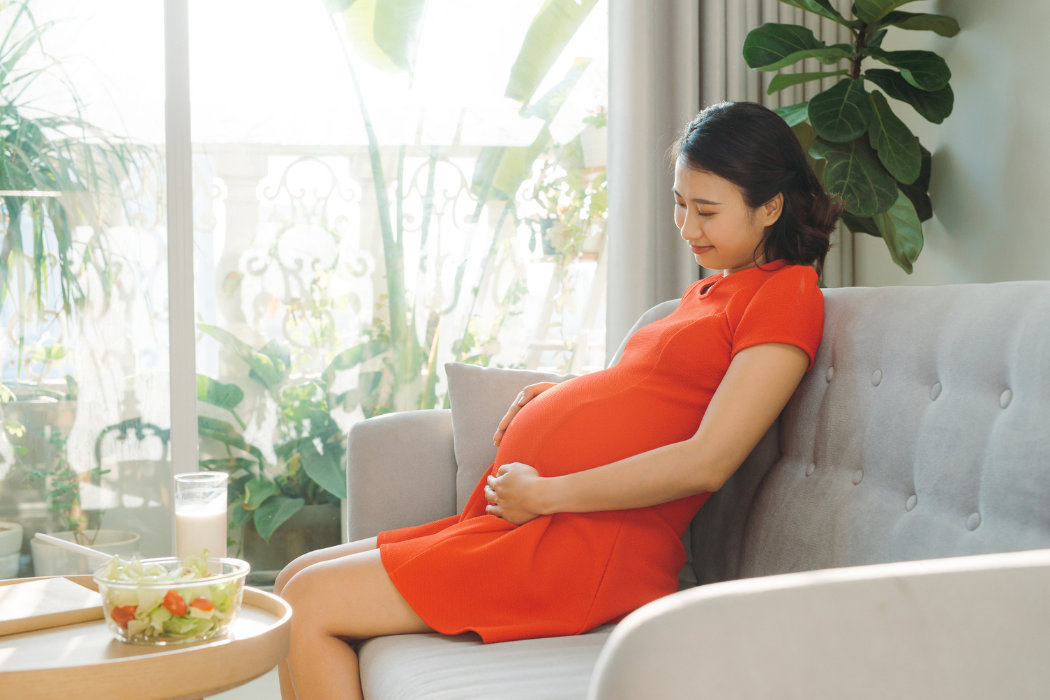
Uterine fibroids are quite common in women of the reproductive age group and as such, commonly encountered in pregnancy.
Though majority of these cases are asymptomatic, some are prone to developing complications and may end up having adverse outcomes in pregnancy.
Let’s explore more on uterine fibroids and pregnancy with Dr Tricia Ruth Thomas, who specialises in Obstetrics and Gynaecology (O&G), from Pantai Hospital Klang.
What are uterine fibroids?

Fibroids or leiomyomas are benign uterine tumours that arise from the myometrium or muscular layer of the uterus.
Typically, there are 3 types, intramural, subserosa or submucosal, located within the body of the uterus. Intramural fibroids grow within the middle and thickest layer of the uterus (called the myometrium). They are the most common fibroids.
Subserous fibroids grow out from the thin outer fibrous layer of the uterus and can hang by a thin stalk attached to the uterus, these are called pedunculate fibroids. Submucous fibroids grow from the uterine wall toward and into the inner lining of the uterus (the endometrium). Fibroids can also present in the cervix or broad ligament.
What are the symptoms of fibroids?
Some fibroids maybe asymptomatic, but others typically cause heavy and or prolonged menstrual bleeding.
Sometimes bleeding can be so heavy that it may result in anaemia.
As fibroids grow larger, they can cause pressure effects on the surrounding structures.
Pressure on the urinary bladder may lead to urgency or frequency of urination or can even cause urinary retention. Pressure on the colon may cause bowel dysfunction and constipation. They may also cause subfertility and recurrent miscarriages.
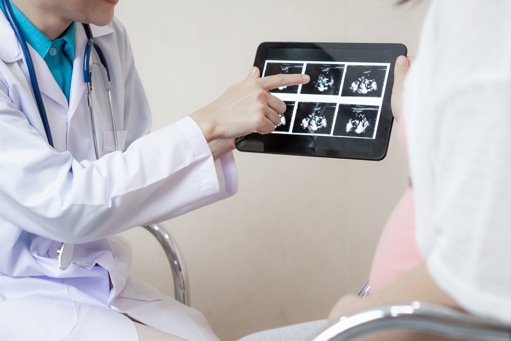
What are the types of tests that we can take to detect uterine fibroids?
Fibroids are usually diagnosed with a bedside abdominal or pelvic ultrasound. However, they can also be assessed by CT scan or MRI (Magnetic Resonance Imaging).
At what age will women have uterine fibroids? Why do fibroids develop at this age?
Typically, fibroids are seen in the reproductive age group, where there are high levels of oestrogen & progesterone hormones.
After menopause, when hormone levels are low, they tend to regress or remain small and dormant. Fibroids affect women most commonly in their 30s and 40s, with incidence increasing with age.
What about the size of these uterine fibroids? What is the most common form of uterine fibroids found amongst Malaysian women? Why?
Size can vary from less than a centimetre(cm) to massive tumours more than 20cm in size. Size is not as important as location of the fibroid. Subserosa fibroids can be asymptomatic, while submucosal fibroids commonly cause heavy bleeding. Fibroids are common in all populations, with incidence as high as 40-70%.
How might fibroids affect fertility and pregnancy?
Subfertility is reported in 5-10% of women with fibroids. Fibroids can cause distortion of the uterine cavity or fallopian tubes, causing tubal blockage or effecting the endometrial lining of the womb inhibiting implantation. It can also create a hyperestrogenic environment in the womb, making it unfavourable for pregnancy. In patients who have infertility with a submucosal fibroid, removal of the fibroid or myomectomy is recommended. Two thirds or women conceive following the myomectomy.
How common are fibroids in women and why is it hard to detect them while pregnant?
During pregnancy, some fibroids remain the same size and are asymptomatic, not interfering with the pregnancy. They can be detected by ultrasound scans in the early stages, but as the pregnancy and foetus grows, it can be difficult to see them when they remain small. However, some fibroids may grow larger during pregnancy and can be seen on routine ultrasound scans and can cause malposition of the foetus.
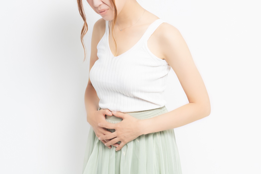
What are the primary problems that could occur from fibroids in first trimester?
In the first trimester, (from 0-12 weeks gestation), fibroids are linked to an increased risk of miscarriage. Some fibroids can undergo degeneration, causing abdominal or pelvic pain.
Risks for pregnant women with fibroids
While some fibroids remain generally asymptomatic during pregnancy, and the pregnancy and delivery go on smoothly without complications, others may cause issues. The rate of complication is higher with larger fibroids, greater than 200 cubic cm, or larger than 5cm. Pain can commonly occur, due to red degeneration of the fibroids, or torsion of a pedunculated fibroid. Other complications include preterm labour and placental abruption which is the premature separation of the placenta from the uterine wall. This is especially high with submucosal fibroids located adjacent to the placental site. The risk of placenta previa (low lying placenta) is also higher with fibroids. In addition, larger fibroids can distort the uterine cavity causing malpresentation or malposition of the foetus. Fibroids located within the lower uterine segment are associated with higher Caesarean section rates and retained placentas.
Can fibroids cause complications during delivery?
There is an increased risk of bleeding or postpartum haemorrhage with fibroids, as they interfere with the contractility of the uterus after delivery of the baby. Hence, in these instances, women are advised to deliver in high risk units with facilities for blood transfusion and Caesarean section option.
Do fibroids go away after pregnancy?
Fibroids usually do not go away after delivery, but they may shrink in size and become less detectable on scans. They are found to regress in over 70% cases and become asymptomatic. However, regular follow up to monitor the progress of the fibroids is recommended.
What is the most common treatment for fibroids?

Treatment for fibroids is usually surgical, both open and laparoscopic removal possible depending on the size and location of the fibroids.
Myomectomies are done for women who are keen to retain their uterus, whilst hysterectomies are performed in older women who have no future childbearing wishes.
Women with previous myomectomies, with the endometrial cavity breached, or with large fibroids, are usually advised to deliver by Caesarean section, to reduce risk of uterine rupture.
A non-surgical treatment modality is uterine artery embolization, an interventional radiological procedure. However, this is associated with higher risk of miscarriages and placenta accrete (morbidly adherent placenta) in subsequent pregnancies.
During pregnancy, fibroids are usually managed conservatively with no active intervention till after delivery and the postnatal period.